An 11 year old boy came into our practice after a fall on his hand and arm. He had swelling and pain with moving his elbow and had lost mobility. His mom needed advice to decide what to do. Her son did not have pain unless he moved it and she thought he might have gotten better on his own, but his pain persisted.
Diagnostic ultrasound is a fast screening tool that we can use in minutes to determine what course of action needs to be done. In this case the ultrasound gave us much more information than the x-ray.
Diagnostic ultrasound using the Butterfly iQ is a fast way to gain in-depth information of soft tissues and even the cortex of bone. Clinical findings of swelling to the right elbow, pain with palpation, and loss of mobility with pain with any movement were present.
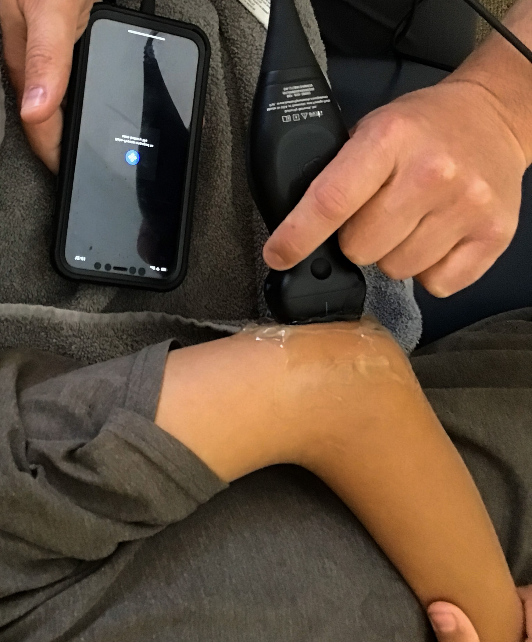
This is the orientation of the ultrasound probe on the elbow.


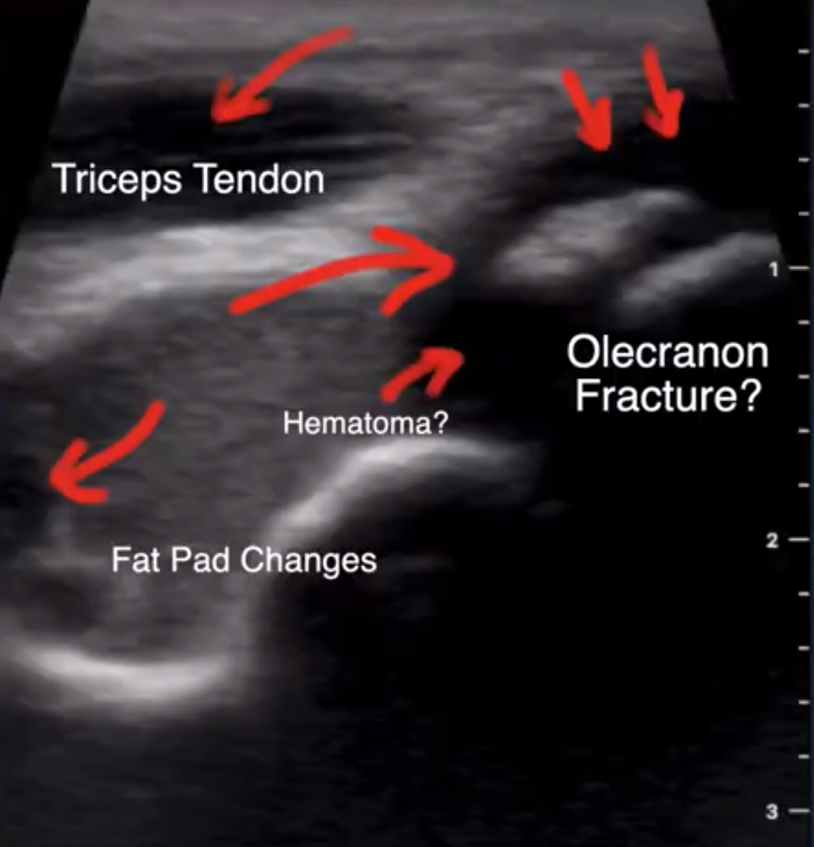
The ultrasound showed an abnormal signal to the fat pad as well as to the tip of the olecranon. The hypo-echoic or darker area around the olecranon is consistent with fluid but not a normal signal for this area. There is a piece of bone (large arrow), or hyper-echoic area, in the center of the hypo-echoic area. You can see lines through this fragment. Based on the history of the injury and the ultrasound imaging a referral to orthopedic surgery was recommended.
ORTHOPEDIC SURGERY CONSULT
Multiple x-rays showed that there 'may be a fracture'. His orthopedic surgeon was unsure. There were suspicions of changes to the right elbow x-rays but they were not conclusive. Bracing increased elbow pain, and stiffness was getting worse.
PHYSICAL THERAPY FOLLOW UP
We repeated the ultrasound and added the left side for comparison. Both elbows showed a hypo-echoic area surrounding a hyper-echoic area consistent with a bone fragment at the tip of the olecranon. This would most likely be a congenital abnormality and not a fracture. I learned that even though the ultrasound finding is abnormal it may be normal for that person. It is easy to image the other side for comparison. This example showed that this boy injured his right elbow but it was not fractured and letting him slowly progress on his own with physical therapy to restore mobility was the course of action needed.
Diagnostic ultrasound was very clear in showing changes to the elbow and should be considered as a frontline screening tool for musculoskeletal injuries.
For more on diagnostic ultrasound and rotator cuff injuries check out this link.